
Retinal Detachment Surgery:
Types, Recovery, Success Rate and Cost

- Retinal detachment is an eye emergency that needs prompt assessment and treatment.
- The type of surgery depends on where and how the retina has detached.
- Treatment may include laser or cryotherapy, pneumatic retinopexy, scleral buckle or vitrectomy.
- Recovery takes time. Clear vision may not return immediately, even when the retina has been successfully reattached.
- The macula plays an important role in visual recovery.
- Retinal reattachment is successful in around 9 out of 10 cases, although some patients may need another procedure.
- The cost depends on the treatment required and the complexity of the retinal detachment.
Seeing flashes of light. Suddenly noticing many more floaters. A dark shadow that seems to be moving across your vision.
These symptoms can be frightening, especially when you are told that you may have a retinal detachment.
The questions usually come quickly:
“Will I need a retinal detachment surgery?”
“Will my vision come back?”
“How long will it take to recover?”
A retinal detachment needs to be assessed promptly. The longer the retina remains detached, the greater the concern for permanent damage to vision. In many cases, surgery is needed to bring the retina back into position and treat the problem that caused it.
But there is something important to understand.
There is no single retinal detachment surgery that is right for everyone.
The location and extent of the detachment, the retinal tear, whether the macula is involved, and whether there is scar tissue or traction all influence the treatment we choose.
Let’s understand what that means.
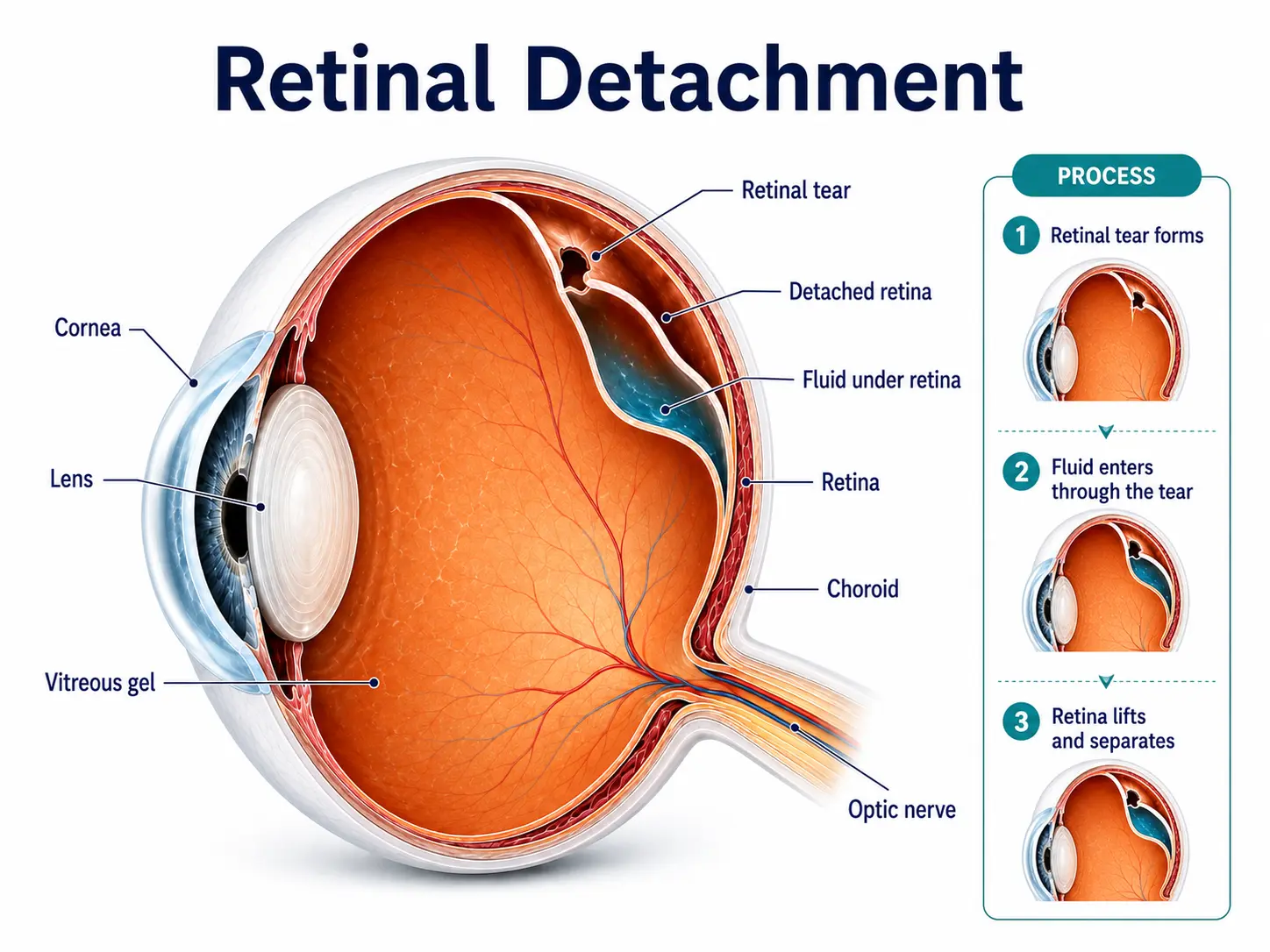
What Exactly Is a Retinal Detachment?
The retina is the light-sensitive layer at the back of your eye. It receives light and sends signals to the brain, allowing you to see.
It needs to stay in its normal position to work properly.
A retinal detachment occurs when the retina separates from the tissue supporting it. When this happens, the retina cannot function normally, and vision can be affected.
Sometimes, a tear in the retina allows fluid to pass underneath it and lift it away.
In other cases, scar tissue or traction pulls the retina away from where it should be.
The extent of the detachment matters too.
If the macula; the part of the retina responsible for detailed central vision, is still attached, the situation can be different from one in which the macula has already detached.
What Are the Early Warning Signs?
Yes. OCP is more than an eye disease.
The same type of tissue affected in the eye also lines several other parts of the body. Symptoms may therefore appear in one area first and another area months or even years later.
Seek an urgent retinal examination if you suddenly notice:
- Flashes of light
- A sudden increase in floaters
- Blurred or reduced vision
- A curtain or shadow across your vision
- Distorted vision
- Sudden loss of part or all of your vision
A few floaters do not automatically mean you have a retinal detachment. Floaters can occur for several reasons.
What matters is a sudden change.
Especially when flashes, a sudden shower of floaters or a curtain-like shadow appear together.
In that situation, we would much rather examine your retina and tell you that everything is fine than have you wait at home and risk delaying treatment.

Why Does Early Treatment Matter?
Once a retinal detachment is diagnosed, timing matters.
The longer the retina remains detached, the greater the risk of lasting damage to vision. An early examination can also tell us whether the macula is still attached and how much of the retina is involved.
Sometimes a retinal tear is found before the retina has detached.
In those situations, laser treatment or cryotherapy may be enough to seal the tear and prevent a detachment from developing.
That is why sudden flashes, new floaters, a curtain-like shadow or sudden vision loss should never be ignored.
The sooner the retina is examined, the sooner the right treatment can begin.
Does Every Retinal Detachment Need the Same Surgery?
No.
This is one of the most important things to understand.
We don’t choose a procedure simply because someone has been diagnosed with retinal detachment.
We first look at what is actually happening inside the eye.
We consider:
- Where the retinal tears are
- How large the detachment is
- Whether the macula is attached
- Whether there is scar tissue or traction
- The condition of the lens
- Whether there is bleeding or another retinal problem
- Previous eye surgery or trauma
For some retinal breaks, laser or cryotherapy may be sufficient.
When the retina has already detached, treatment may involve pneumatic retinopexy, scleral buckle surgery, vitrectomy or a combination of procedures.
The aim is the same: reattach and support the retina while addressing the underlying problem.
What Are the Different Types of Retinal Detachment Surgery?
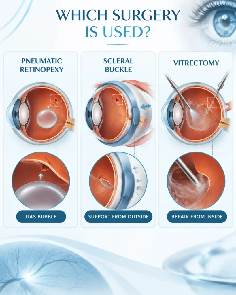
Pneumatic Retinopexy
Pneumatic retinopexy may be suitable for selected retinal detachments, depending on the location and characteristics of the tear.
During the procedure, a gas bubble is placed inside the eye. The bubble rises and presses against the detached retina, helping bring it back into position. Laser treatment or cryotherapy is then used to seal the retinal tear.
One part of recovery is particularly important here: head positioning.
Depending on where the tear is located, you may need to keep your head in a particular position so the gas bubble stays against the area being treated.
There are also important precautions while the gas remains in the eye.
Flying and travelling to high altitudes should be avoided. Nitrous oxide anaesthesia must also be avoided because it can cause the gas bubble to expand and dangerously increase eye pressure.
Your ophthalmologist will tell you when these restrictions can safely be lifted.
Scleral Buckle Surgery
A scleral buckle takes a different approach.
Instead of supporting the retina from inside the eye, we support it from the outside.
A silicone band is placed around the outer wall of the eye. This creates a gentle indentation, bringing the area around the retinal tear closer to the detached retina and helping support the repair. Laser or cryotherapy may also be used.
The buckle is generally left in place.
Some redness, watering or discomfort can occur while the eye heals. Some patients may also notice a change in their spectacle prescription because the shape of the eye can change slightly after surgery.
Vitrectomy
Vitrectomy is commonly used when the retinal detachment is more complex.
For example, it may be recommended when there are multiple tears, significant traction or scar tissue, bleeding inside the eye, or a more extensive detachment.
During the procedure, the vitreous gel inside the eye is removed through very small openings.
This allows the surgeon to access the retina, relieve traction, treat retinal tears and reposition the detached retina.
The eye may then be filled with air, gas or silicone oil to support the retina while it heals.
In more complicated cases, vitrectomy may be combined with a scleral buckle.
How Long Does Retinal Detachment Surgery Take?
“Doctor, how long will my surgery take?”
It is one of the first practical questions patients ask.
There isn't one fixed retinal detachment surgery time.
A straightforward repair may be relatively quick. A more complicated case involving multiple tears, scar tissue or additional procedures can take longer.
A vitrectomy may take around 1–3 hours, depending on the complexity of the case.
But try not to judge the severity of your retinal detachment by the length of the operation.
A longer surgery does not automatically mean a worse prognosis.
What matters more is the condition of the retina, whether the macula is involved and how effectively the underlying problem can be repaired.
What Is Recovery Like After Retinal Detachment Surgery?
This is where we like to set realistic expectations from the beginning.
The surgery repairs the retina. It does not mean your vision will become clear immediately.
In the early stages, the eye may be red, watery or uncomfortable. Vision can also be blurry, particularly when a gas bubble has been placed inside the eye.
Recovery is gradual.
Depending on the procedure and the condition of the retina, recovery may take around 4–8 weeks, while more complex vitrectomy cases can take 6–12 weeks.
During recovery, you may be asked to:
Your individual recovery may be different.
So don't judge your final vision from the first few days or weeks.
We monitor the retina during follow-up because healing is a process.
“Doctor, the Retina Is Attached. Why Is My Vision Still Blurred?”
This is one of the most worrying things patients notice after surgery.
They may come for a follow-up and ask:
“You said the retina is attached. Why can't I see properly yet?”
The reason is that retinal reattachment and visual recovery are not the same thing.
If the macula was detached, the cells responsible for detailed central vision may already have been affected before surgery. Even after the retina is successfully reattached, vision can take time to recover, and it may not always return to its previous level.
A gas bubble or silicone oil can also temporarily make vision appear blurred.
So blurred or distorted vision after retinal detachment surgery does not automatically mean the surgery has failed.
During follow-up, we look at the retina and the overall recovery rather than judging the outcome only by how clear your vision feels in the early stages.
What Is the Retinal Detachment Surgery Success Rate?
We understand why patients want a number.
Based on the hospital's reference information, successful retinal reattachment occurs in approximately 9 out of 10 patients, although some patients may require another procedure.
But there is an important distinction.
A successfully reattached retina does not guarantee completely normal vision.
Your visual prognosis depends significantly on the condition of the retina before surgery, particularly whether the macula was involved.
Can the Retina Detach Again After Surgery?
Yes.
A retinal detachment can recur in some patients even after an initially successful repair.
That is why follow-up remains important.
When you return for your post-operative examinations, we are not simply checking whether the eye looks better. We are checking the retina, looking for new or recurrent retinal problems and monitoring how your recovery is progressing.
Contact your eye specialist promptly if you notice:
Don’t assume that a sudden change is simply part of normal recovery.
When Should You See a Retina Specialist?
If you suddenly notice flashes, a shower of new floaters, a curtain-like shadow, blurred vision or sudden vision loss, don’t wait for pain. Retinal detachment can be painless and needs urgent assessment.
During an examination, we check the retina to understand where the problem is, how extensive it is and whether the macula is involved. If there is a retinal tear or detachment, treatment is chosen based on these findings and may include laser, cryotherapy or surgery.
If surgery is needed, we explain what to expect, including the procedure, recovery, visual outcome and likely cost.
Retinal detachment is serious, but it is treatable. Surgery can reattach the retina, although visual recovery takes time and may be incomplete, particularly when the macula is affected.
If you notice sudden flashes, new floaters, a curtain or shadow, or vision loss, seek an urgent retinal examination.