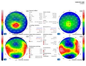
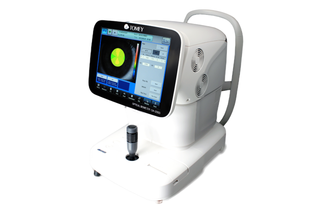
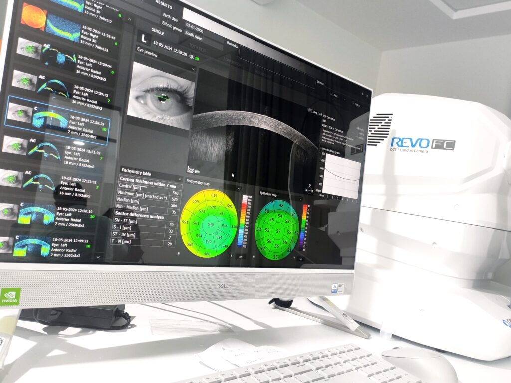
Our corneal topography and tomography systems provide precise mapping of corneal curvature, thickness, and shape, facilitating early detection of conditions such as keratoconus and guiding refractive surgery planning. These advanced technologies ensure accurate assessment and personalized treatment recommendations for optimal visual outcomes.
Our refractive surgery screening evaluates candidacy for procedures such as LASIK and PRK, assessing corneal thickness, curvature, and refractive error to determine suitability and predict post-operative outcomes. With our advanced diagnostic tools, we ensure safe and effective treatment for refractive errors.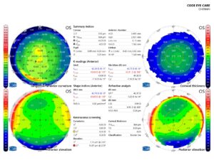

Our corneal topography and tomography systems provide precise mapping of corneal curvature, thickness, and shape, facilitating early detection of conditions such as keratoconus and guiding refractive surgery planning. These advanced technologies ensure accurate assessment and personalized treatment recommendations for optimal visual outcomes.
Utilizing corneal topography and tomography data, we offer comprehensive keratoconus screening to detect subtle changes in corneal morphology indicative of this progressive condition. Early identification allows for timely intervention to slow disease progression and preserve visual function.
Our refractive surgery screening evaluates candidacy for procedures such as LASIK and PRK, assessing corneal thickness, curvature, and refractive error to determine suitability and predict post-operative outcomes. With our advanced diagnostic tools, we ensure safe and effective treatment for refractive errors.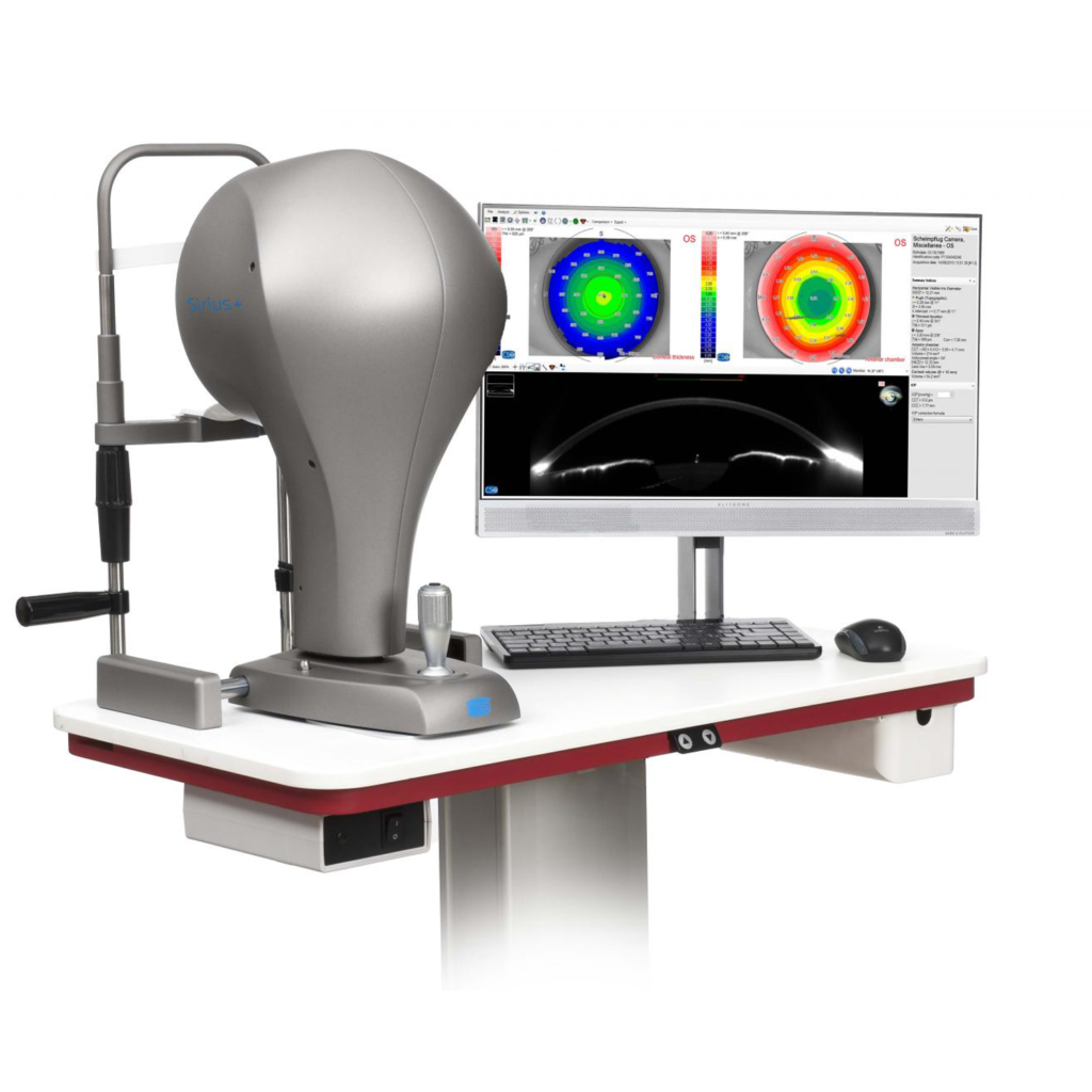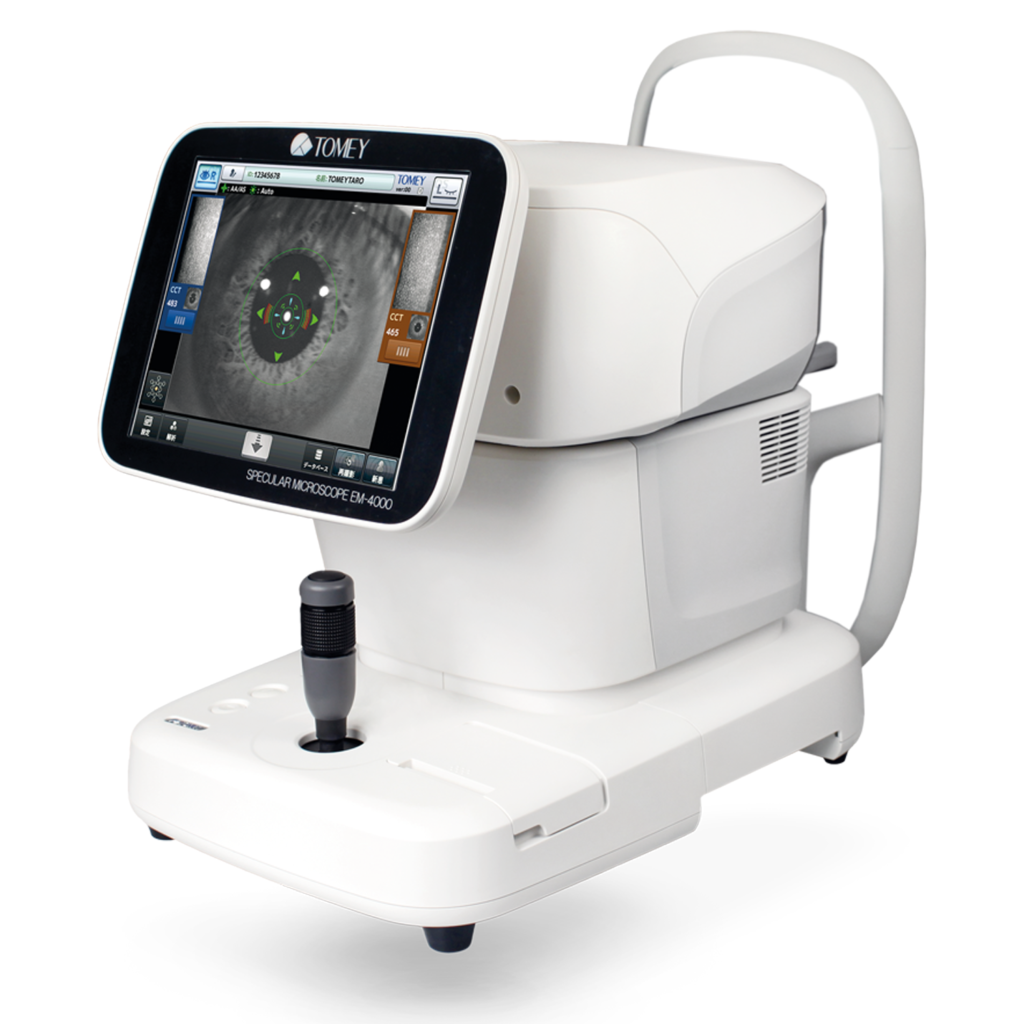
Our corneal topography and tomography systems provide precise mapping of corneal curvature, thickness, and shape, facilitating early detection of conditions such as keratoconus and guiding refractive surgery planning. These advanced technologies ensure accurate assessment and personalized treatment recommendations for optimal visual
outcomes.
Utilizing corneal topography and tomography data, we offer comprehensive keratoconus screening to detect subtle changes in corneal morphology indicative of this progressive condition. Early identification allows for timely intervention to slow disease progression and preserve visual function.
Our refractive surgery screening evaluates candidacy for procedures such as LASIK and PRK, assessing corneal thickness, curvature, and refractive error to determine suitability and predict post-operative outcomes. With our advanced diagnostic tools, we ensure safe and effective treatment for refractive errors.
Our IOL calculation services utilize advanced biometry techniques and sophisticated formulas to ensure accurate selection of intraocular lenses for cataract surgery and refractive lens exchange. With precise measurements of axial length, corneal power, and anterior chamber depth, we tailor IOL selection to each patient’s unique ocular anatomy and visual needs.
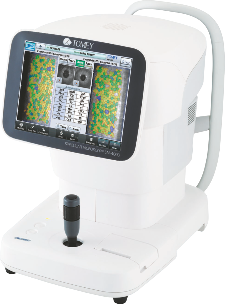
Autorefraction provides objective measurements of refractive error, allowing for rapid and accurate assessment of the eye’s optical properties. This non-invasive technique serves as a valuable starting point for determining spectacle prescriptions and guiding further refractive evaluations.
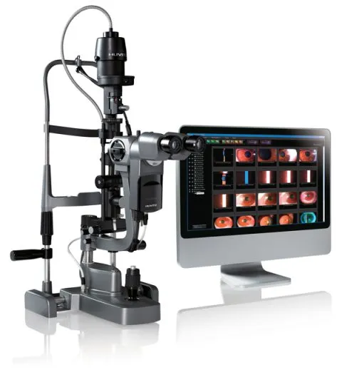
Comprehensive refraction services fine-tune spectacle prescriptions through subjective assessment of visual acuity, binocular vision, and accommodative function. Our experienced optometrists utilize a combination of traditional techniques and advanced technologies to optimize visual performance and comfort for patients of all ages.